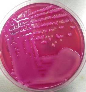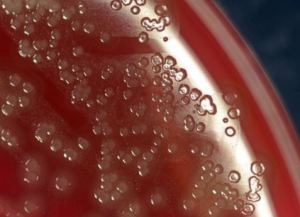
Bacteria grow on solid media as colonies. A colony is defined as a visible mass of microorganisms originating from a single mother cell. Key features of these bacterial colonies serve as important criteria for their identification.

Colony morphology can sometimes be useful in bacterial identification. Colonies are described based on size, shape, texture, elevation, pigmentation, and effect on growth medium.
Find common criteria that are used to characterize bacterial growth;
Table of Contents
It includes the form, elevation, and margin of the bacterial colony.
Form of the bacterial colony: The form refers to the shape of the colony. These four forms represent the most common colony shapes you are likely to encounter.

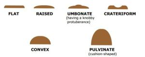
Elevation of the bacterial colony: It gives information about how much the colony rises above the agar. This describes the “side view” of a colony.
 Elevation of Bacteria colony" width="" height="" />
The six most common elevations of bacterial colonies are
Colonies that are irregular in shape and/or have irregular margins are likely to be motile organisms. Highly motile organisms swarmed over the culture media, such as Proteus spp.

The size of the colony can be a useful characteristic for identification. The diameter of a representative colony may be measured in millimeters or described in relative terms such as pinpoint, small, medium, and large.
Tiny colonies are also referred to as punctiform (pin-point). Colonies larger than about 5 mm are likely to be motile organisms. Punctiform colonies are distinguished from circular colonies by their very small size.

Bacterial colonies are frequently shiny and smooth in appearance. Other surface descriptions might be: dull (opposite of glistening), veined, rough, wrinkled (or shriveled), or glistening. Bacillus species give dry, wrinkled colonies. Pseudomonas stutzeri also gives similar-appearing wrinkled colonies.
Several terms that may be appropriate for describing the texture or consistency of bacterial growth are: dry, moist, viscid (sticks to loop, hard to get off), brittle/friable (dry, breaks apart), mucoid (sticky, mucus-like).
Some bacteria produce pigment when they grow in the medium, e.g., green pigment produces by Pseudomonas aeruginosa, buff-colored colonies of Mycobacterium tuberculosis in L.J medium, and red-colored colonies of Serratia marcescens.
The opacity of a bacterial colony can be described as transparent (clear), opaque (not transparent or clear), translucent (almost clear, but distorted vision–like looking through frosted glass), or iridescent (changing colors in reflected light). A pinpoint translucent β-hemolytic colonies on blood agar is most probably a Streptococcus species. Staphylococci give opaque, smooth, and circular colonies on the agar plate surface.
Draughtsman colonies

Young colonies of Streptococcus pneumoniae (pneumococci) have raised centers, but as the culture ages, they become flattened, with a depressed central part and raised edges giving them a ringed appearance also known as ‘draughtsman colonies’.